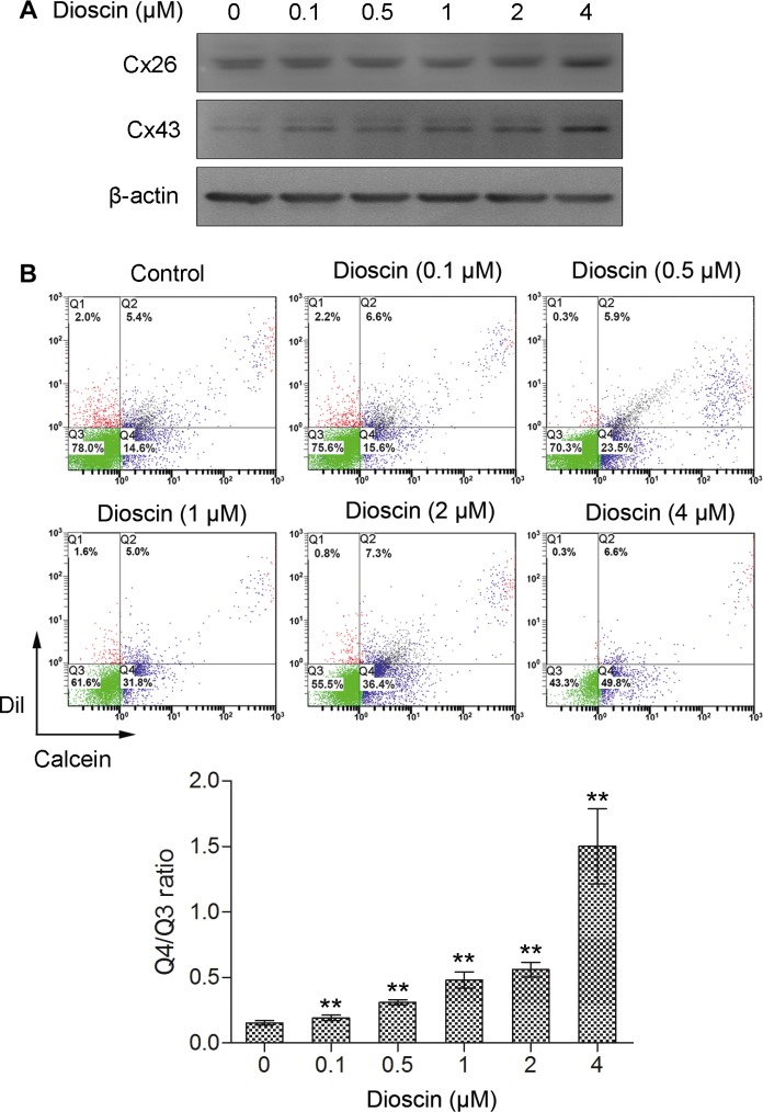
Figure 2. Increase of GJIC by dioscin in B16 melanoma cells.
(A) Upregulation of Cx26 and Cx43 proteins in dioscin-treated B16 cells examined by immunoblotting (B) Promotion of GJIC by dioscin in B16 cells, as measured by fluorescent dye transfer assay. Q2: DiI and Calcein double-positive cell populations (donor cells); Q4: Calcein-positive cells (recipient cells). The ratio of the B16 cell number in Q4 to that in Q3 (double negative cells) was used to evaluate GJIC function. The lower panel shows the quantification from three independent experiments. **P < 0.01, compared with control.

