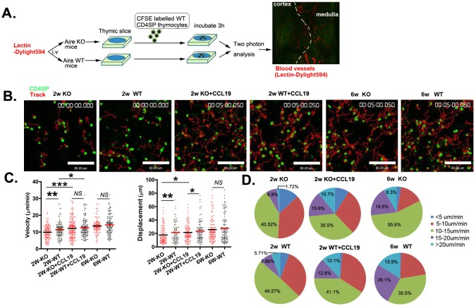
Figure 5. The reduced motility of CD4SP thymocytes within the thymic tissue of neonatal Aire KO mice.
A. The experimental design used to analyse CD4SP thymocyte migration in sliced thymic tissues using a two-photon microscope. Dylight594 conjugated lectin was injected to the Aire−/− and Aire+/+ mice just prior to sacrifice. 4×10⁵ CFSE-labelled CD4SP cells were loaded onto thymic slices from Aire−/− or Aire+/+ mice, and the cells on the slice were incubated for 3h at 37°C/5% CO2 to allow cells to enter the tissue. For CCL19 rescue assay, CD4SP cells were incubated with 200ng/ml CCL19 and then loaded on the thymic tissues. The slice was imaged by two photon microscope. The thymic structures were visualized by the labelled blood vessels. B. The movements of individual cells were tracked. CD4SP (green). Tracks (red). Scale bar, 80 μm. C. The average velocity of CD4SP in the medulla are shown in left panel. Bars indicate the median. Average displacements from the origin plotted are also shown in right panel. The raw tracking data for CD4SP thymocytes are from three independent experiments. (2 weeks-old Aire−/−, n = 116, Aire+/+ mice, n = 140; 2 weeks-old with CCL19 Aire−/−, n = 168 and Aire+/+ mice,n = 140, 6 weeks-old Aire−/−,n = 108 and Aire+/+ mice, n = 138). D. Profiles of the migration velocities of CD4SP cells on different thymic slices. p values were calculated by Mann-Whitney U-test, * p < 0.05, ** p < 0.01, *** p < 0.001. NS, no significance.

