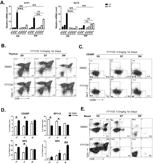
Figure 6. The role of S1P1 signal in neonatal thymic output.
A. The four subgroups of CD4SP thymocytes from 2 and 6-week old Aire−/− mice and littermates were sorted. The mRNA expression levels of S1P1 and KLF2 were analyzed by qPCR. GAPDH was used as internal standard. B.-E. FTY720 (0.5mg/kg) was administrated to wild type mice from D5 to 2 weeks old by intraperitoneally injection for 2 days. B.-D. 24 hours later, thymocytes were collected for CD4,CD8,CD69 and Qa-2 staining. The ratio of CD4SP in total thymocytes and SP1 to SP4 cells in CD4SP were indicated as mean± SD. * p < 0.05. ** p < 0.01. E. Blood depleted red cells was collected for CD4 and CD8 staining. Representative dot plots were shown. N = 5. * p < 0.05, ** p < 0.01, *** p < 0.001. NS, no significance.

