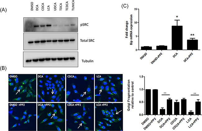
Figure 4. Unconjugated bile acids cause Golgi fragmentation and Bip activation via activation of Src.

A. HET-1A cells were treated with Bile acids and phosphorylation of Src was measured by western blot. B. Cells were pre-treated with PP2 (10 μM), prior to treatment with bile acids. Golgi structure was assessed using a GM130 antibody (green) and imaged using confocal microscopy as outlined in materials and methods. Nuclei were stained with Hoechst (blue). C. HET-1A cells were pre-treated with PP2 (10 μM) followed by treatment with DCA. Levels of Bip mRNA were quantified by RT-PCR. Data are represented as mean ± SEM, normalised to vehicle control for n=3 experiments, * p<0.05 versus Control **p<0.05 versus bile acid without PP2.
