. 2017 Mar 15;6:e20882. doi: 10.7554/eLife.20882

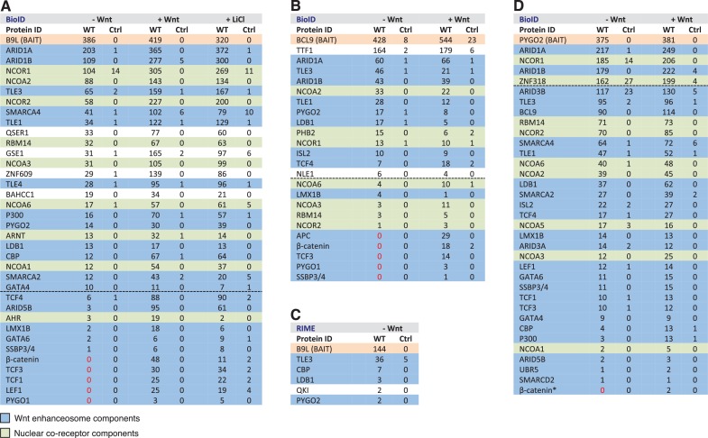
Figure 4. BCL9/B9L and PYGO2 are constitutively associated with the Wnt enhanceosome, and nuclear co-receptor complexes.

(A, B) List of BioID hits for (A) B9L-BirA* and (B) BCL9-BirA*±10–12 hr of WCM; names above the dotted line refer to the top hits, while names below this line refer to hits selected on relevance to Wnt (blue) or nuclear co-receptors (green); only specific hits with a > 5 spectral count ratio relative to the BirA* control are shown; numbers represent unweighted spectral counts (>95% probability). (C) RIME hits for FLAG-B9L-BirA*; only specific hits with a >5 spectral count ratio relative to the control are shown. (D) List of BioID hits for PYGO2-BirA*, as in (A, B); *, identified with lower confidence (>55% probability).

DOI: http://dx.doi.org/10.7554/eLife.20882.009

Figure 4.

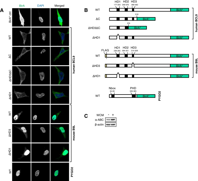
Figure 4—figure supplement 1. Stably transfected BCL9/B9L cell lines for BioID, and summary of wt and mutant BirA* baits.

Figure 4—figure supplement 1.

(A) Immunofluorescence of stable BioID T-REx-293 cell lines expressing wt or mutant BCL9-BirA*, B9L- BirA* or BirA* alone, showing subcellular distribution of the various baits; nuclei are labeled by DAPI staining. (B) Cartoons of wt and mutant BCL9-BirA*, B9L-BirA* and PYGO2-BirA*. (C) Western blot probed with a-ABC antibody, to confirm Wnt induction of the stimulated PYGO2-BirA* cell lysates prior to BioID pull-downs and analysis.
Figure 4—figure supplement 2. Additional analysis of BioID hits.
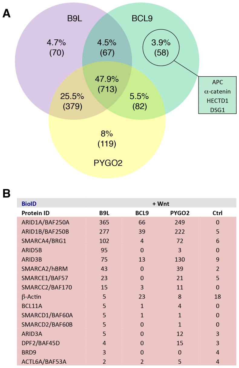
Figure 4—figure supplement 2.

(A) Venn diagrams, showing the number of hits shared between the BCL9, B9L and PYGO2 BioID baits with >95% probability (excluding the BirA*-only control);right, a selection of high-confidence BCL9-specific hits not found with the other two (nuclear) baits. (B) Components of the SWI/SNF complex found by all three baits.
Figure 4—figure supplement 3. Constitutive association between B9L and TCF prior to Wnt stimulation.
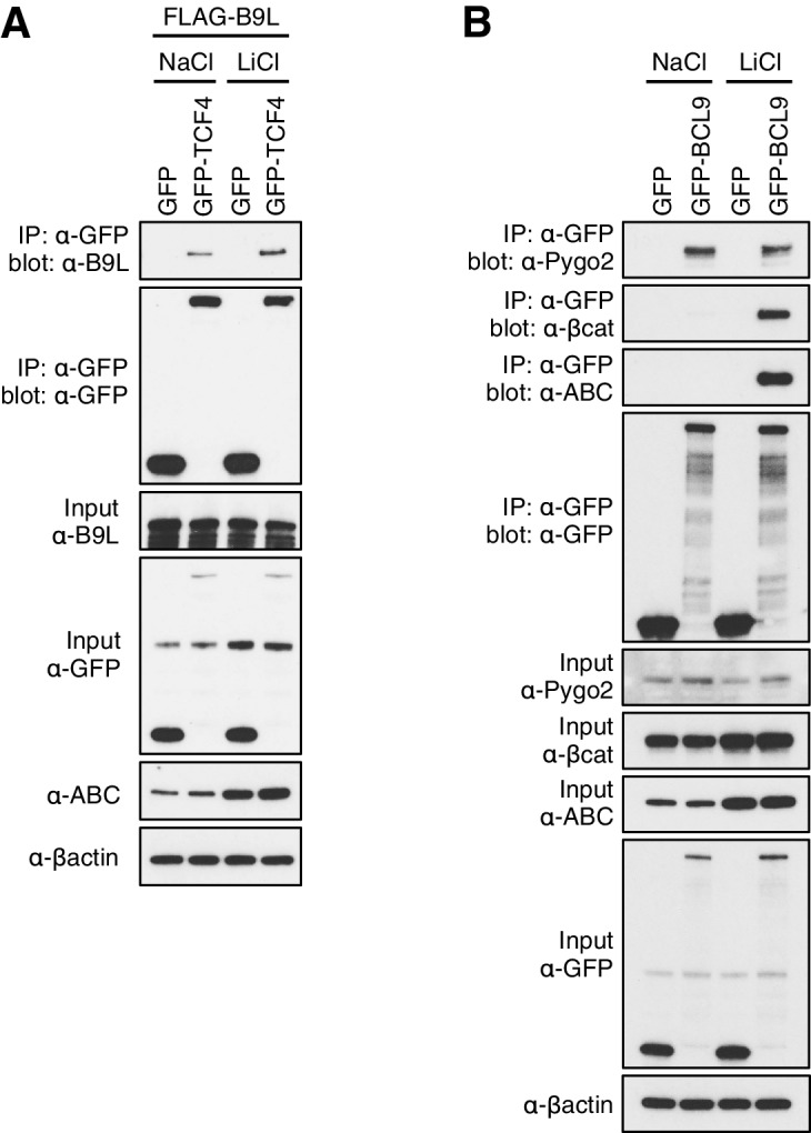
Figure 4—figure supplement 3.

CoIP assays in transiently transfected HEK293T cell, with Western blots showing (A) Wnt-independent association between GFP-TCF and FLAG-B9L, or (B) Wnt-dependent association between GFP-BCL9 with endogenous b-catenin or ABC (and Wnt-independent association with endogenous Pygo, as internal control). Transfected cells were exposed to LiCl, or NaCl as control, as described in the main Materials and methods.