
Figure 6. Variant viral transcription during early stages of B-cell infection by B95-8/npcEBNA1.
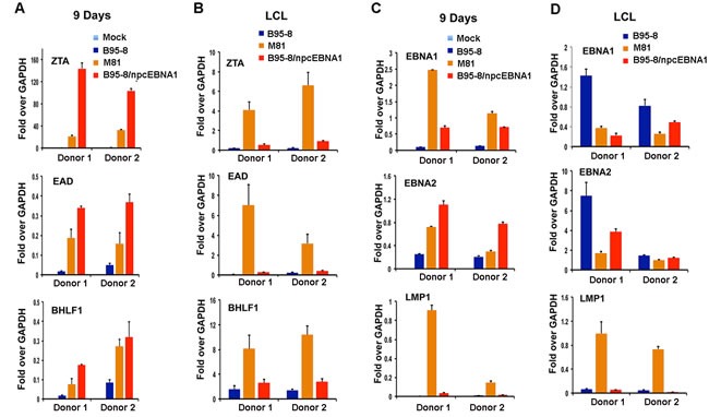
RT-PCR of primary B-cells infected with either mock, or bacmid derived B95-8, M81, or B95-8/npcEBNA1 virus at either 9 days post-infection (panels A and C) or for established LCLs at 2 months post-infection (panels B and D). EBV lytic transcripts for ZTA, EA-D, or BHLF1 (panels A and B), or latency associated transcripts for EBNA1, EBNA2, or LMP1 (panels C and D) are shown relative to GAPDH. RT-PCR is shown as the average for three replicates for two independent donors. Error bars represent standard deviation from the mean.