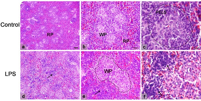
Figure 6. Histological structure of the chicken spleen.

a.-c. The normal spleen structure. The red and white pulps are clearly distinguished. d.-f. The spleen structure after LPS injection. Lymphocytes irregularly presented in the red pulp (arrow), but disappeared in the PELS of white pulp. WP, white pulp; RP, red pulp; PELS, periellipsoidal lymphatic sheath.
