
Figure 1. Work flow of the sample preparation for mass spectrometry (MS) analysis.
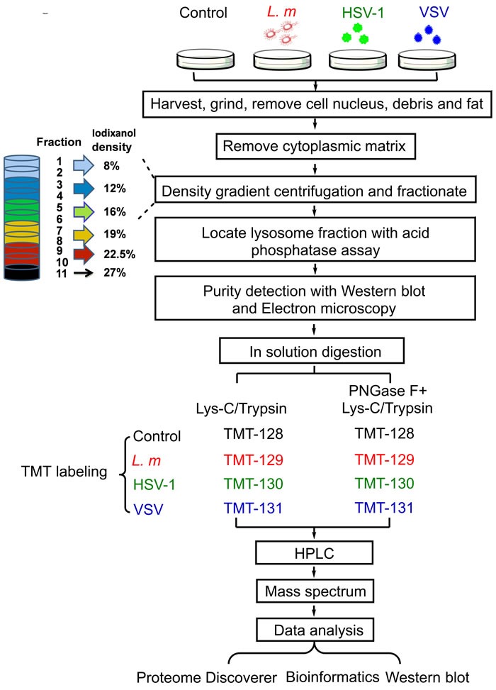
Mouse macrophage cell line RAW 264.7 were infected with L. m, HSV-1 and VSV or culture medium for 9 hours followed by harvest and purification. Lysosomes were purified by density gradient centrifugation and fractionation. Lysosome purity was detected with western blot and electron microscopy. High purity lysosomes were used in subsequent TMT based quantitative MS and analysis.