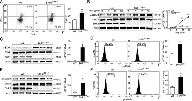
Figure 4. SHP2-deficiency in CD4+ T cells enhances IFN-γ-STAT1 signaling and Th1 differentiation.

(A) Flow cytometry analysis of CD4+IFN-γ+ cells in the spleen from WT and SHP2CD4−/− mice. (B) Immunoblotting analysis of STAT1 activation in CD4+ T cells from WT and SHP2CD4−/− mice in the presence of 20 ng/ml IFN-γ for indicated time. (C, D) Immunoblotting analysis (C) or flow cytometry analysis (D) of STAT1 activation in CD4+ T cells from mesenteric lymph node from DSS-induced colitis mice at day 8. (E, F) Immunoblotting analysis (E) or flow cytometry analysis (F) of STAT1 activation in CD4+ T cells from mesenteric lymph node from AOM-DSS-induced CAC mice at day 80. Data are representative of three independent experiments (mean ± SEM of 6 mice per group). *P < 0.05, **P < 0.01 vs. WT group.
