
Figure 3. The relationship between the expression of STAT3 and lymph node metastasis.
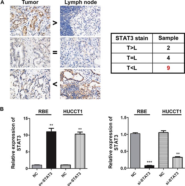
(A) The expression levels of STAT3 in ICC and relevant lymph nodes were detected by Immunohistochemical test (n = 15). Representative image is illustrated. (B) The STAT3 expressions were detected by real-time PCR 2 days after over expression or inhibition of it in RBE and HUCCT1 cells, respectively. **P < 0.01, ***P < 0.001, compared with negative control group.