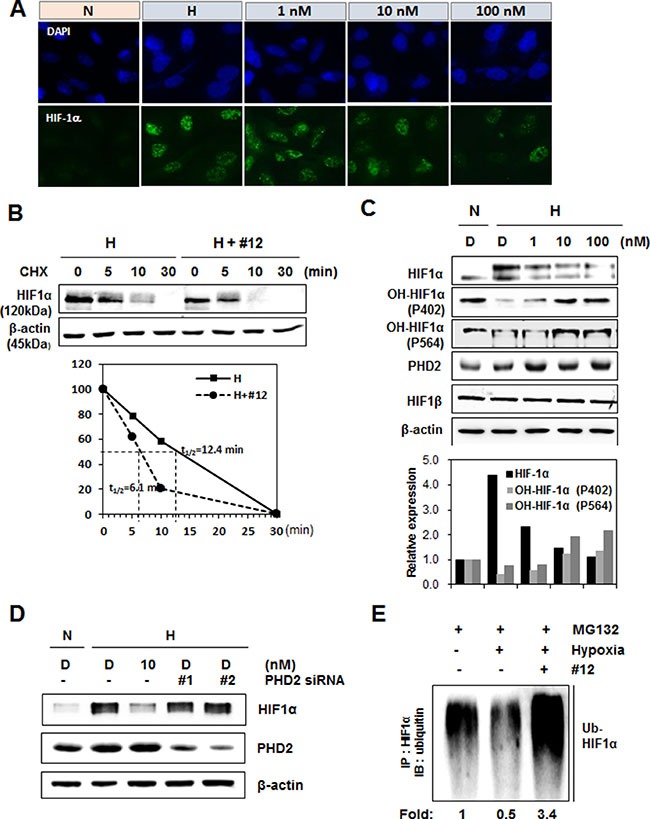
Figure 4. Compound 12 increased hydroxylation and proteasome-mediated degradation of HIF-1α.

(A) A549 cells were treated with Compound 12 (at concentrations of 0 nM, 1 nM, 10 nM, and 100 nM) for 16 h. Cells were fixed with paraformaldehyde; an immunofluorescence assay was then performed using anti-HIF-1α antibodies and Alexa Fluor 488-labeled secondary antibodies (green). Nuclei were stained with DAPI. N, normoxia; H, hypoxia. (B) A549 cells were treated with Compound 12 (10 nM concentration) and exposed to hypoxic conditions for 16 h. Cells were immediately treated with cycloheximide (CHX, 100 μg/mL) for the time indicated, and HIF-1α protein levels were determined by western blotting. β-actin was used as an internal control. Half-life (t1/2) of HIF-1α protein was determined using relative expression levels of HIF-1α. (C) A549 cells were treated with Compound 12 (at concentrations of 0 nM, 1 nM, 10 nM, and 100 nM) for 16 h. The expression levels of HIF-1α, hydroxylated (OH)-HIF-1α at P402 or P564, and PHD2 were determined via western blotting using specific antibodies. N, normoxia; H, hypoxia. (D) A549 cells were transfected with two kinds of small interfering RNAs targeting PHD2 and then treated with Compound 12 under hypoxic exposure for 16 h. Cell lysates were immunoblotted with an anti-HIF-1α and PHD2 antibody. (E) After A549 cells were treated with Compound 12 for 16 h, cells were treated with MG132 (20 μM) for 4 h. Cell lysates were immunoprecipitated (IP) with anti-HIF-1α antibody, and were then immunoblotted (IB) with anti-ubiquitin antibody. Relative levels of ubiquitinated HIF-1α were quantified and graphed.
