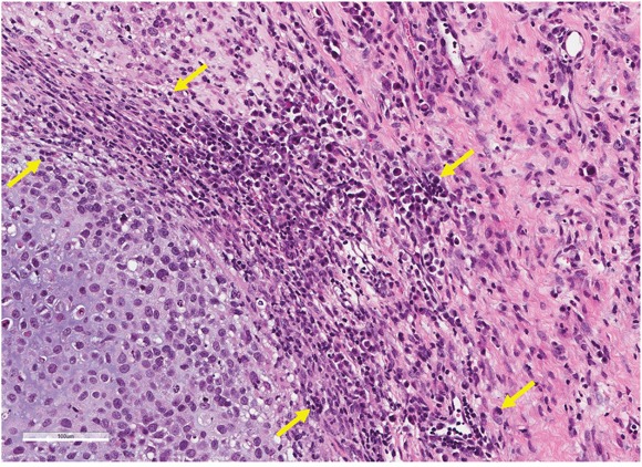
Figure 4. Effect of S. typhimurium on tumor histology.

Tumors were resected from nude mice at autospy, fixed in formalin, embedded in paraffin, sectioned and stained with hematoxylin and eosin (H&E) by standard methods. The figure shows the histology of an osteosarcoma treated with S. typhimurium A1-R. Necrotic areas are indicated by yellow arrows. Scale bar: 100 μm.
