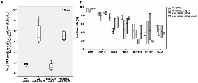
Figure 4. In vivo assays.

A. Percentage of GFP-positive cells in the peripheral blood of lethally irradiated recipient mice transplanted with PML/RARA-positive or WT bone marrow cells in the presence or absence of ΔNp73 overexpression. B. Immunophenotypic analysis of bone marrow recipients’ cells with respect to myeloid and lymphoid cell markers after transplantation. Box plots show the summarized data for immunophenotypic analyses of bone marrow from survival mice that received a transplant of empty vector control (WT, 7 animals; hCG-PML/RARA, 8 animals) or ΔNp73 (WT, 8 animals; hCG-PML/RARA, 6 animals). hGC-PML/RARA transplanted mice were not leukemic at the time of analysis. Bone marrow cells were stained for the indicated surface markers as indicated in the bottom of the figure. No significant difference between groups were detected.
