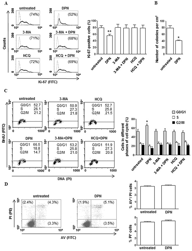
Figure 1. DPN reduces cell proliferation and alters cell cycle progression in HL cells.

A. Cell proliferation was evaluated by flow cytometry measuring Ki-67 nuclear antigen expression in L-428 cells treated or not with 10 nM DPN for 48 hours in the presence or absence of 3-MA or HCQ. Results from one representative experiment out of 3 are shown (left). Data are also reported as mean ± SD (right), **, p < 0.01 versus untreated cells. B. The effects of DPN (10 nM) on L-428 long-term survival was determined by in vitro colony formation assay. The mean (± SD) values correspond to three independent experiments, *, p < 0.05. C. Cell cycle progression was evaluated by flow cytometry using the BrdU/anti-BrdU method in synchronized L-428 cells treated or not with 10 nM DPN for 48 hours in the presence or absence of 3-MA or HCQ. Results from one representative experiment out of 3 are shown (left). Data are also reported as mean ± SD (right), *, p < 0.05 versus untreated cells. D. Apoptosis/necrosis assay involving dual staining with AV and PI was carried out using flow cytometry in L-428 cells treated or not with 10 nM DPN for 48 hours. Results from one representative experiment out of 3 are shown (left). Numbers reported represent the percentages of AV positive/PI negative (early apoptotic, bottom right quadrant) and PI positive (late apoptotic or necrotic cells, top right and left quadrants). Data are also reported as mean ± SD (right).
