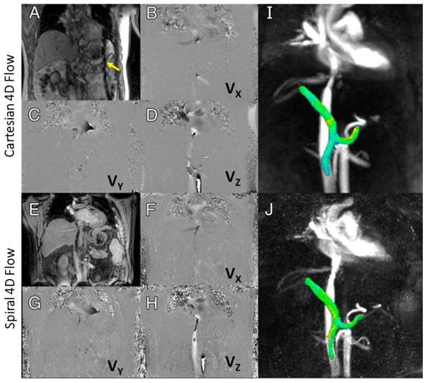
Figure 12.
Representative results obtained with Cartesian (A–D, I) and spiral (E–H, J) 4D flow imaging techniques. Magnitude images are shown in A and E and phase-difference images are shown in B–D and F–H. VX, VY, and VZ correspond to velocity measured with motion-encoding gradients in right-left, anteriorposterior, and foot-head directions, respectively. I and J are 3D angiograms showing segmented view of portal, splenic, and superior mesenteric veins with comparable quality and conspicuity. Dark lines proximal to spleen on Cartesian series (yellow arrow) show cross-beam navigator used for respiratory gating in Cartesian acquisition. (Images were obtained from the Figure 2 in Dyvorne H et al. Radiology 2015 Apr;275(1):245–54 and were reproduced with permission from the authors and the journal.)

