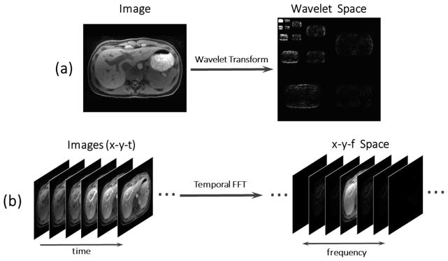
Figure 2.
MR images can be considered sparse in an appropriate transform domain. A liver image has as parse representation (i.e. a representation with a small number of high-value coefficients) in the wavelet space (a), and a dynamic contrast-enhanced image series has a sparse representation in the x-y-f (two spatial dimensions + temporal frequency dimension) space, with a FFT (fast Fourier transform) performed along the temporal dimension (b).

