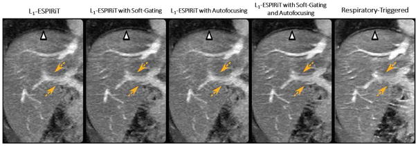
Figure 7.
L1-ESPIRiT with either soft-gating or autofocusing achieved better image quality of the portal vein than L1-ESPIRiT without motion compensation (yellow dashed arrows). A combination of soft-gating and autofocusing enabled further improvement in delineation of the liver dome (white arrow) and the hepatic vessels (yellow dashed arrows) compared to L1-ESPIRiT with soft-gating or autofocusing alone, and also achieved better delineation of the hepatic vessels than the respiratory triggered reference. (Images were obtained from the Figure 4 in Cheng JY et al. J Magn Reson Imaging. 2015 2015 Aug;42(2):407–20 and were reproduced with permission from the authors and the journal.)

