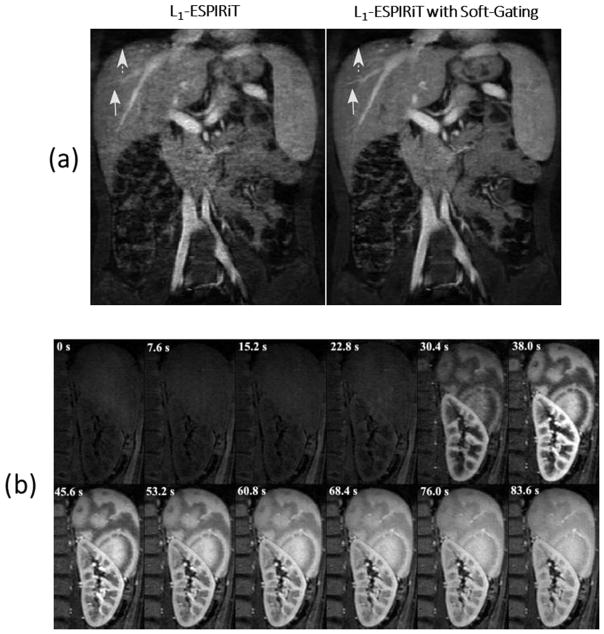
Figure 9.
(a) L1-ESPIRiT reconstruction with no motion weighting (left) and with soft respiratory gating (right). Soft-gating improves the delineation of the liver edge (dashed arrows) and the hepatic vessels (solid arrows). (b) Zoomed and cropped image of the spleen and kidney at different contrast enhancement phases with a spatial resolution of 1.1x1.1 mm2. The time of acquisition is shown on top of each contrast phase. Images show the progressive enhancement from cortical to medullary region of the kidney, as well as the perfusion pattern of the spleen. (Images were obtained from the Figure 2c and Figure 5a in Zhang T et al. J Magn Reson Imaging. 2015 Feb;41(2):460–73 and were reproduced with permission from the authors and the journal.)

