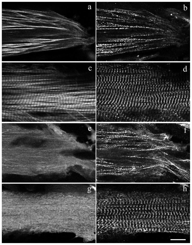
Figure 12. Fluorescence image of quail myotubes transfected with YFP fused zebrafish and TPM1-2ν.
Skeletal muscle myotubes were transfected with YFP fused zebrafish TPM1-2α (a,c) and TPM1-2ν (e,g). Cells were then fixed and stained with alpha-actinin antibody to localize premyofibrils at the spreading end of myotubes (b,f) and mature myofibrils in the central regions of myotubes (d,h). Note that the YFP-TPM1-2α localized in both premyofibrils (a,b) and mature myofibrils (c,d), while YFP-TPM1-2ν is in the mostly diffused area in cells with very weak localization in the myofibrils (e–h). The expression of YFP-TPM1-2ν does not affect myofibrils in the myotube (f,h). Bar = 10 μm.

