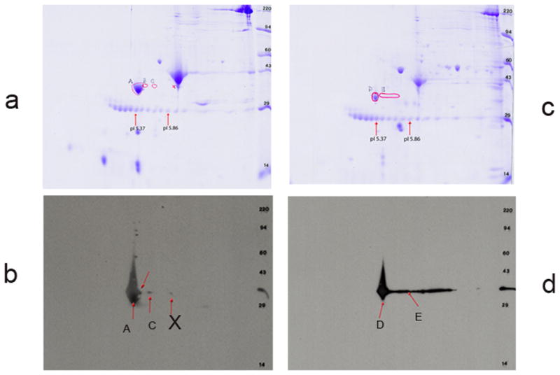
Figure 13. 2D western blot analyses with extracts from adult zebrafish Heart and skeletal muscle.

- The Coomassie stained zebrafish skeletal muscle protein across the gel.
- The PVDF filter was stained with CH1 monoclonal antibody followed by treatment with secondary antibody as mentioned under materials and methods, and subsequently treated with ECL and exposed to x-ray film. Developed X-ray film was superimposed on the top of the Coomassie stained second gel as well as on the Coomassie stained PVDF filter. Three spots A, C, X were marked, excised and was used for extraction of protein for subsequent Mass spectrometric analyses.
- The Coomassie stained zebrafish cardiac muscle protein across the gel.
- Same as in b. However, in this gel 2 spots D & E were marked for subsequent Mass Spectrometric analyses.
