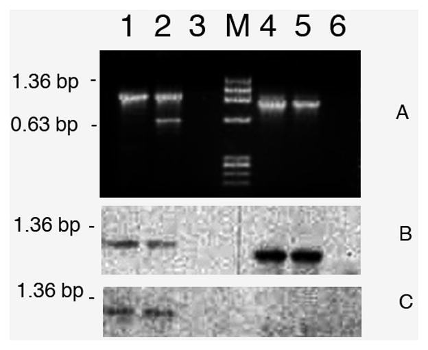
Figure 2. Conventional RT-PCR amplification of TPM1α and TPM1κ with RNA from heart and skeletal muscle of Zebrafish.

Lane 1. Amplification of both TPM1α and TPM1κ by generic primer-pair (Gen TPM1-1α&κ(+)/Gen TPM1-1α&κ(−) as given in Table 1) in heart; lane 2. Amplification of both TPM1α and TPM1κ by generic primer-pair in sk muscle; lane 3. Primer control; lane M. Marker; lane 4. Amplification of TPM1κ by isoform specific primer pair (TPM1-1κ Primer (+)/Gen TPM1-1α&κ(−) as given in Table 1) in heart; lane 5. Amplification of TPM1κ in skeletal muscle; lane 6. Primer control.
