
Figure 4. Conventional RT-PCR amplification of TPM4-1α and total TPM4-1 transcripts expressed in zebrafish heart and skeletal muscle.
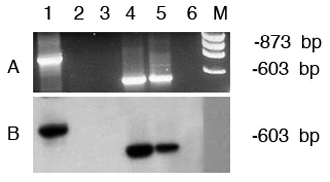
- Ethidium bromide staining of the agarose gel
- Hybridization with TPM4-1-specific probe
Lane 1.TPM4α in heart; lane 2. TPM4α in skeletal muscle; lane 3. Primer control; lane 4. Total TPM4-1 in heart; lane 5. Total TPM4-1 in skeletal muscle; lane 6. Primer control.
For lane 1to 3 PCR amplification was carried out with TPM4-1α specific primer-pair as shown in Table 1. The nucleotide sequences of the primer-pair used for the amplification of total TPM4-1 transcripts are also given in Table 1. Please note that the expression of TPM4-1α is practically undetectable in skeletal muscle (lane 2 in panels A and B). However, total TPM4 transcripts are present in both heart and skeletal muscle.