
Figure 1. (A–E). Whole body PET-CT and histopathological features of a lymph node biopsy specimen involved by chronic lymphocytic leukemia (CLL) and necrosis attributable to herpes simplex virus (HSV) infection.
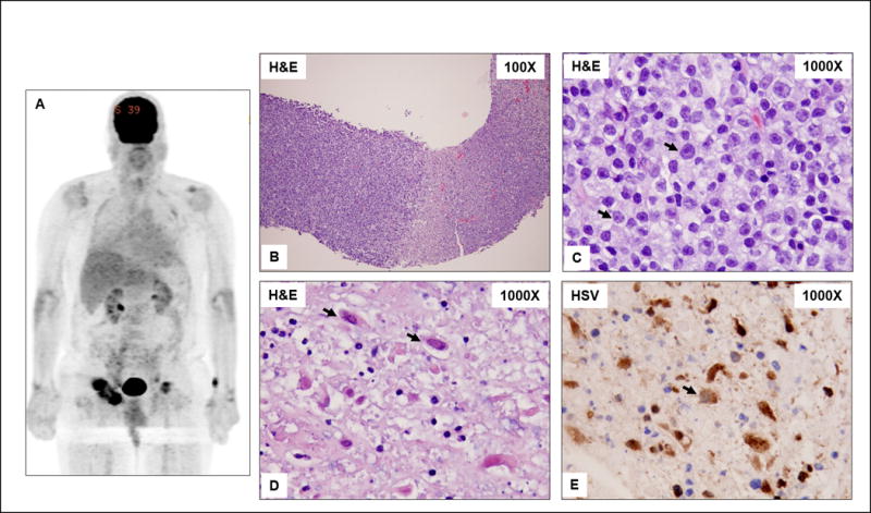
A) Enlarged, metabolically active, likely centrally necrotic, right greater and left inguinal lymph nodes. Prominent 4 × 5 cm right inguinal lymph node with SUV of 9.85 is seen. B) A core needle biopsy specimen showing replacement of lymph node by viable CLL (left of field) and necrosis (right of field). C) High magnification of CLL showing small lymphocytes and increased intermediate-size to large cells (arrows) consistent with prolymphocytes and paraimmunoblasts. D) High magnification of the necrosis showing scattered necrotic large cells with intranuclear inclusions typical of the Cowdry type A HSV inclusions (arrows). E) Immunohistochemistry for HSV (types I and II) is positive in the large cells with viral inclusions (arrows).