
Figure 5.
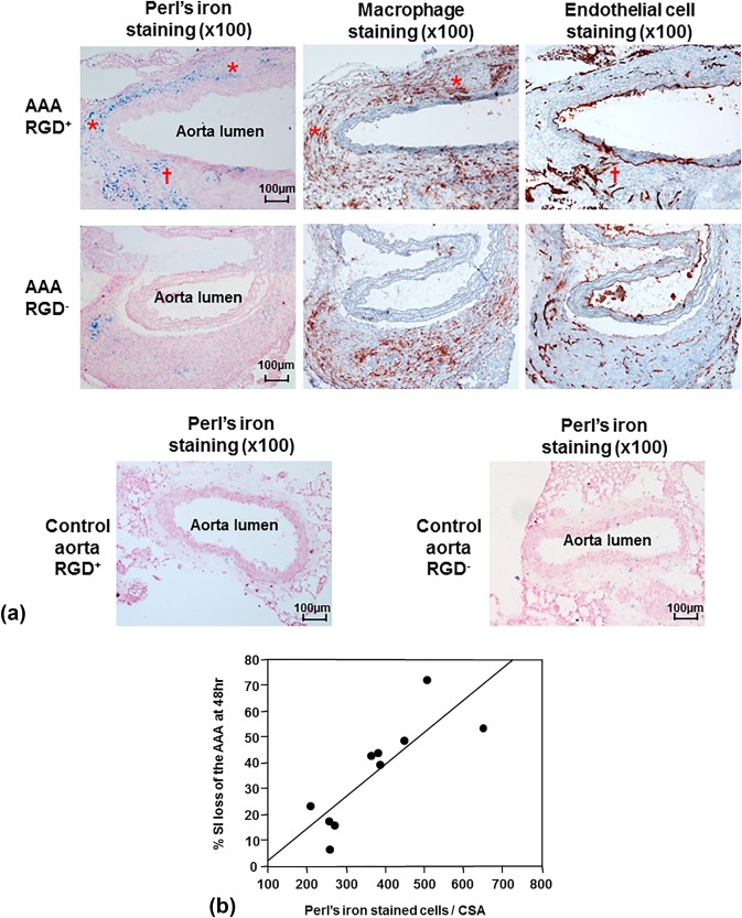
Histological analysis of abdominal aortic aneurysms (AAA) and correlation with signal loss on MRI. A: Immunohistochemical AAA staining showed mural macrophage infiltration and endothelial cell expression within the AAA wall. Perl's iron staining showed greater accumulation of RGD‐HFn‐Fe3O4 in the media and adventitia of AAA wall (AAA RGD+) compared to HFn‐Fe3O4 (AAA RGD–), colocalizing with both macrophages (asterisks) and endothelial cells (dagger). The control aortic wall showed minimal Perl's iron staining in both RGD+ and RGD– groups. B: There was a close correlation between the total number of Perl's iron‐stained cells and % SI loss in the AAA (n = 10, r = 0.83, P = 0.003).