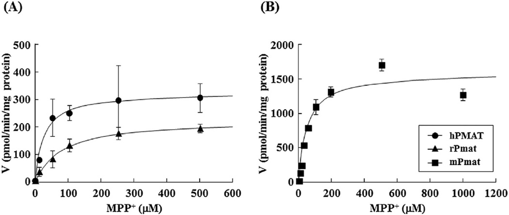
Fig. 3. Concentration dependence of hPMAT-, rPmat-, and mPmat-mediated uptake of MPP+.
(A) The hPMAT- (closed circles) and rPmat- (closed triangles) mediated uptake of MPP+ was measured for 3 min at 37 °C and pH 7.4. (B) The mPMAT- (closed squares) mediated uptake of MPP+ was measured for 2 min at 37 °C and pH 7.4. Cells were incubated with 0.5 mL of KRH buffer containing 0.1 µCi of [3H]MPP+ and various concentrations of unlabeled MPP+. The hPMAT- and rPmat-mediated uptake was determined by subtracting the uptake by MDCK/mock cells from that by MDCK/hPMAT and MDCK/rPmat cells, respectively. The mPmat-mediated uptake was determined by subtracting the uptake by Flp293/mock cells from that by Flp293/mPmat cells. Data are shown as the mean ± SEM (n = 3).

