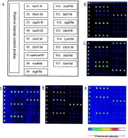
FIG. 1.
Layout of probes on biochips and fluorescence images of clinical isolates. (A) Probe layout on the oligonucleotide biochip. Following hybridization of Cy5-labeled fragmented DNA, as described in Materials and Methods, the fluorescence signals of mutations in the gyrA and parC genes from four clinical isolates were detected, as follows: Ser91→Phe/Asp95→Gly/Ser87→Arg (B), Ser-91→Phe/Asp95→Gly/Ser87→Asn/Glu91→Gln (C), Ser91→Phe/Asp95→Ala/Asp86→Asn/Ser87→Ile (D), and Ser91→Phe/Asp95→Asn/Glu91→Lys (E). (F) Wild-type sequence in gyrA and parC (Ser91/Asp95/Asp86/Ser87/Glu91).

