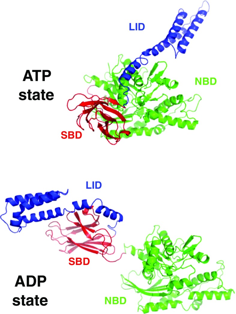
Fig. 1.
Structures of the E. coli Hsp70 protein DnaK in the ADP/NRLLLTG state (bottom, 2KHO) and in the ATP/Apo state (top, 4B9Q). The nucleotide-binding domains (NBDs in green) have the same orientations. The substrate-binding domain (SBD) is in red. Lid domain (blue). DnaK contains another 30 unstructured residues at the C-terminus of the lid (not shown)

