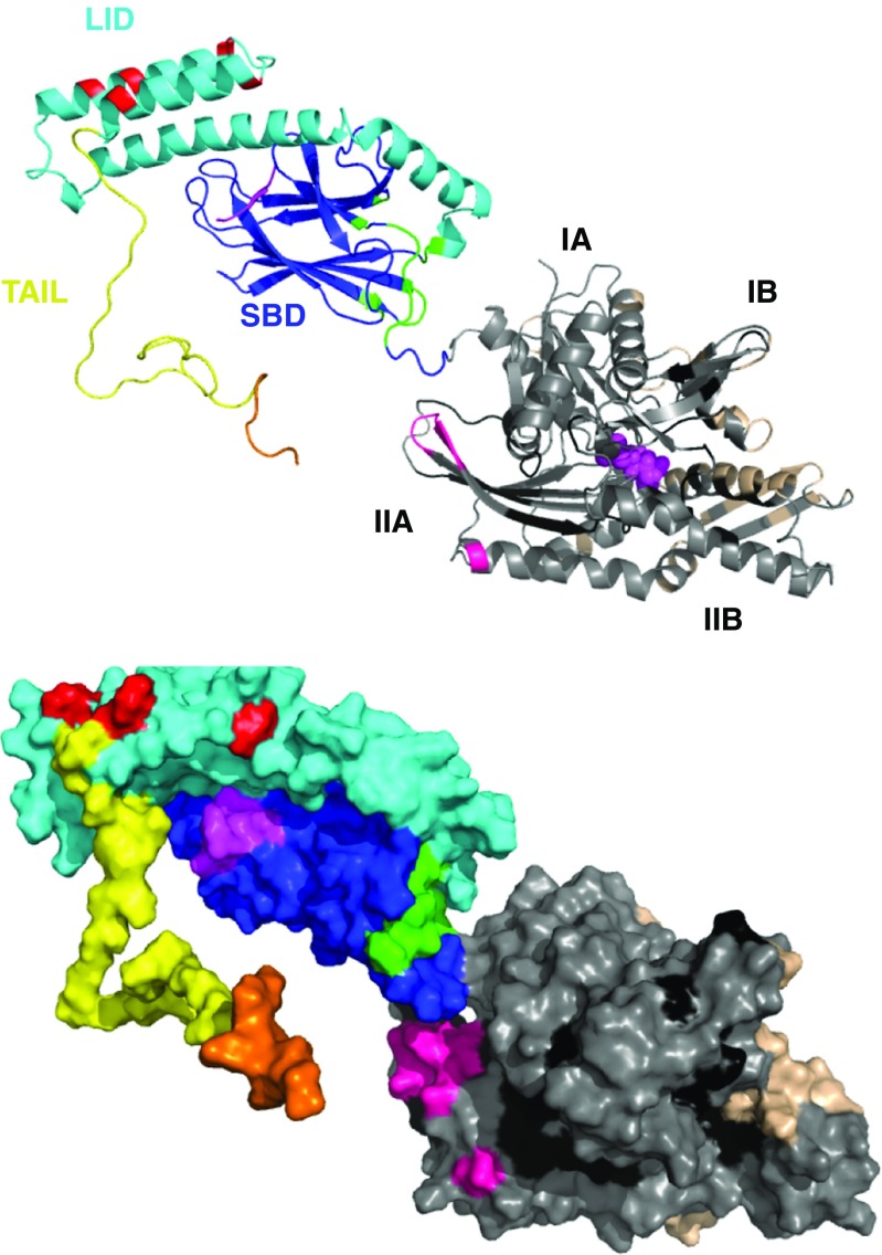
Fig. 9.
Summary of all interaction sites discussed in this review on a homology model for HSPA8 based on DnaK SBD in the ADP state (1DKX), extended with an unstructured tail comprising residues 605–646. NBD, gray; SBD: Blue; LID domain: cyan; tail: yellow. In magenta, substrate peptide NRLLLTG and nucleotide ADP. Interaction sites: all NEFs: beige; all Hsc70 and E. coli DnaK NBD-binding compounds with activity: black. CHIP: orange from (Smith et al. 2013); Phosphoserine lipids: red (Morozova et al. 2016); J-domain – E. coli DnaK interaction from (Ahmad et al. 2011) pink; E. coli DnaK interaction with PET-16 (4R5G): green

