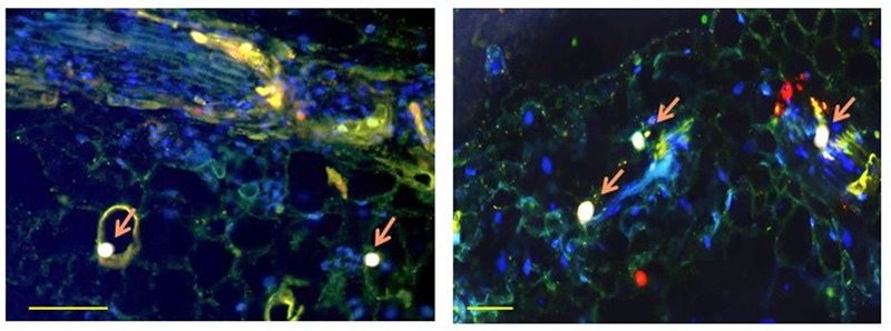
FIGURE 2.

Co-localization of TYLCV CP with tomato HSP70 (left) and with tomato HSP90 (right) in infected leaf at 49 dpi, as observed with a confocal microscope. Cross-section through the leaf blade. CP appears as red, cellular HSP70 or HSP90 as green, nuclei as blue; CP co-localizing with HSP70 or HSP90 in nuclei as pink (pink arrow). Bar is 100 nm. The left photograph is reproduced, with permission, from Gorovits et al. (2013a).
