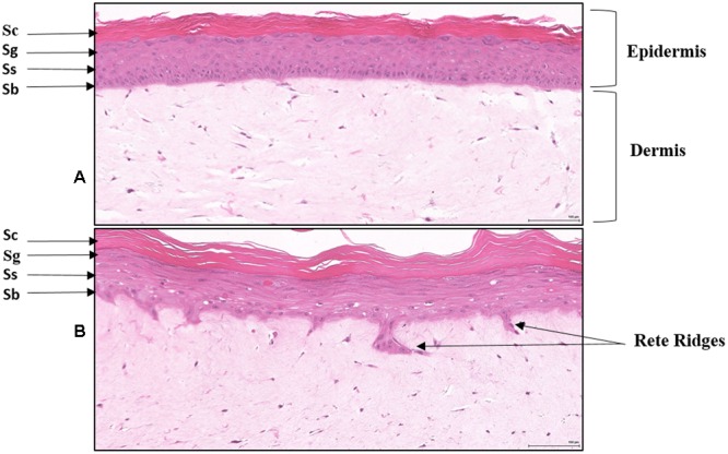
FIGURE 2.

Histology of Reconstituted normal epidermal tissue vs reconstituted psoriatic tissue: H&E stained cross-sections showing reconstituted skin mimics normal epidermis organization and differentiation histology (A). The tissue morphology of the psoriatic tissue model closely parallels that of human psoriatic skin (B). The epidermis contains hyper proliferative basal keratinocytes stratum basale (Sb) with regular elongation of the rete ridges (psoriatic epidermal hyperplasia), and spinous (Ss), granular (Sg), and stratum corneum (Sc) layers. The dermis contains psoriatic fibroblasts (20X). Scale bar = 100 μm.
