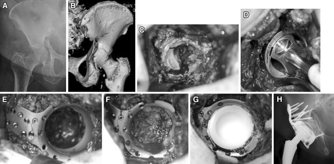
Fig. 2.
a Preoperative a.p. X-ray of a transverse acetabular fracture on the right side in a 79-year-old female. b Posterior view of a 3D-CT scan. c Intraoperative appearance of fracture. d Reaming of the fractured acetabulum up to 52 mm in diameter. e Custom-built Roof-Reinforcement Plate fixed with angle stable 3.5 mm screws. f Bone graft taken from the femoral head padding the cavity of the acetabulum. g Cemented 48 mm polyethylen inlay. h Postoperative a.p. X-ray

