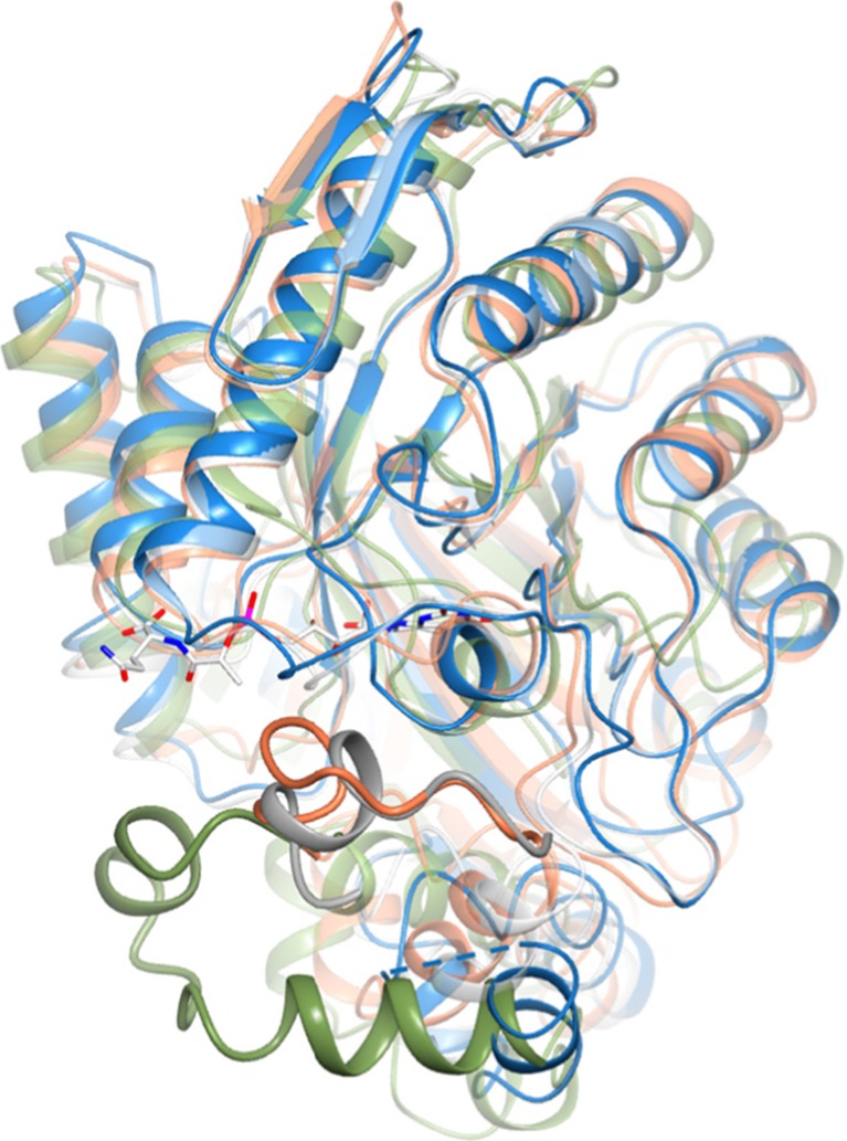
Fig. 6.
Comparison between the active site of Rh-FGD1 (blue) with that of Mtb-FGD1 (3B4Y, white), Adf (1RHC, coral), and Mer (1Z69, green). For clarity, only the F420 from Mtb-FGD1 is shown. The insertion regions of Mtb-FGD1, Adf, and Mer corresponding to the highly disordered segment in Rh-FGD1 (residues 254–263, represented by a dashed line) are highlighted in bold style. The orientation of the molecule is approximately 90° rotated along an axis perpendicular to the plane of the paper with respect to that in Fig. 5c. Color coding for atoms is as in Fig. 5b

