
Figure 2.
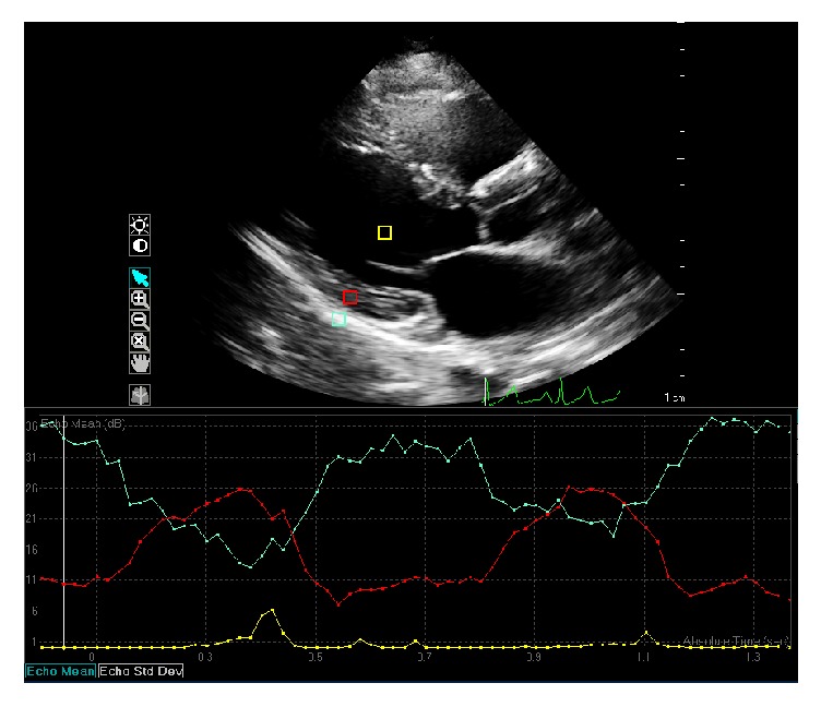
Calibrated integrated backscatter analysis using echocardiography. Region of interest 1 (yellow square) is blood pool within the left ventricle (mean echo-time 0.01 dB). Region of interest 2 (blue square) is the pericardium (mean echo-time 33.96 dB). Region of interest 3 (red square) is myocardium on the posterior wall (mean echo-time 10.29 dB).