
Figure 4.
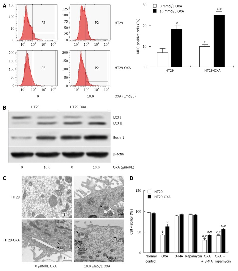
Activation of autophagy contributed to oxaliplatin resistance in colorectal cancer cells. A: HT29 and HT29-oxaliplatin (OXA) cells were incubated with or without 10 μmol/L OXA for 48 h, and autophagy was determined by monodansylcadaverine (MDC)-positive stained cells using flow cytometry; B: Western blotting showed a significant increase in expression of the autophagic markers LC3-II and beclin-1 in HT29 and HT29-OXA cells with or without OXA treatment. The data represent the results of three separate experiments; C: Representative electron micrographs demonstrated autophagic vacuole formation in each group. The arrows indicate the double-membrane vacuoles digesting organelles or cytosolic contents; D: Autophagy regulated chemoresistance of OXA in HT29 and HT29-OXA cells (eP < 0.001 indicates a significant difference vs normal control; aP < 0.05 indicates a significant difference vs 3-MA treatment group; cP < 0.05 indicates a significant difference vs rapamycin treatment group).