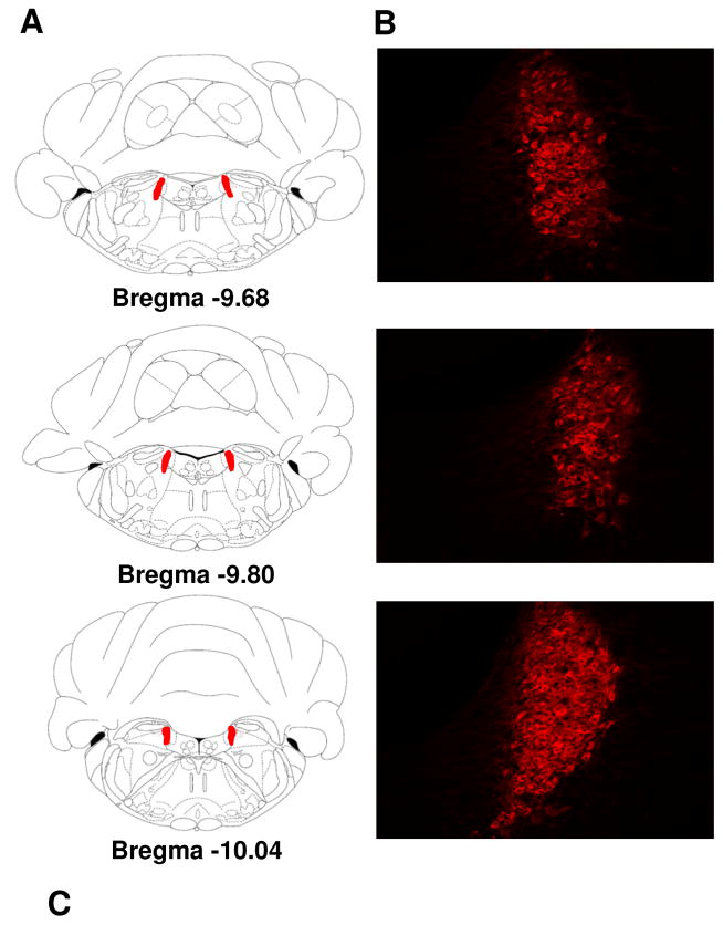
Figure 5.
Alterations in dopamine β hydroxylase (DβH)-positive noradrenergic neurons in the locus coeruleus in air and CO2 exposed Wistar (W), Sprague Dawley (SD), Long Evans (LE) and Wistar Kyoto (WK) rats. (A) Panels on the left show stereotaxic illustrations from the atlas of Paxinos & Watson depicting rostral (bregma −9.68) to caudal (bregma −10.04) from which cells were quantified (red shows LC area). Panel (B) shows representative images showing (DβH) immunopositive cells within the coordinates shown in (A). Panel (C) shows DβH cell counts in air and CO2 exposed animals (* p<0.05 LE and WK air versus SD group; # p= 0.06 LE and WK versus W group; + p<0.05 LE versus WK group). All data are mean ± s.e.m (n=6 animals/air or CO2 groups).

