
Figure 7.
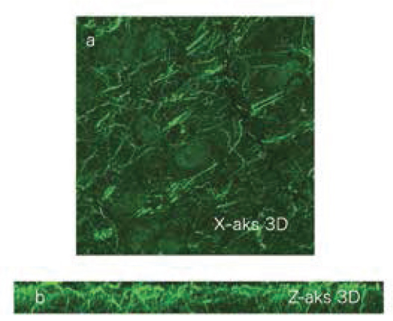
A) On imaging obtained by 3 dimensional overlapping of the sections in Figure 6 in the X axis, filamentous structures are noted clearly. B) Reconstruction of the sections in Figure 6 in the Z axis. The visualized brain tissue has a thickness of 2.2 μm and is an area passing through the striatum. It is noted that the filamentous structure has a netlike construction and extends deeply.