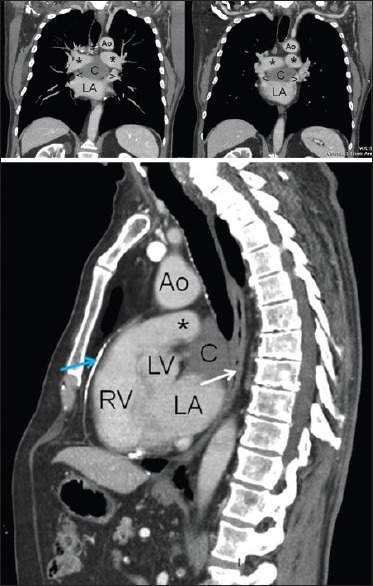
Figure 1.

Thoracic contrast CT scan in coronal (top) and sagittal (bottom) reconstructions. Ao = aorta, C = cyst, LA = left atrium, LV = left ventricle, RV = right ventricle, * = pulmonary arteries, < and > = superior pulmonary veins, white arrow = esophagus, blue arrow = calcified pericardial plaque, CT = computed tomography
