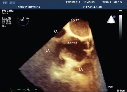
Figure 3.

Three-dimensional TEE at 0°: Intrapericardial cystic mass with moderately echogenic content, well-delineated limits, nonloculated cavity; the walls of the cyst appear thin, smooth, noncalcified, with no signs of echinococcosis; relationships with other structures appear well-defined. LA = left atrium, LV = left ventricle, RA = right atrium, TEE = transesophageal echocardiography
