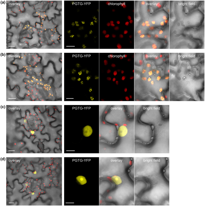
Figure 4. Fluorescence microscopy of YFP-tagged rust effector candidates transiently expressed in tobacco leaves. Confocal images of tobacco cells (Nicotiana tabacum) expressing Puccinia graminis f. sp. tritici (PGTG) effector candidates fused to yellow fluorescent protein (YFP).
Cells expressing PGTG_00164-YFP (a) or PGTG_06076-YFP (b) show chloroplast localisation with a punctate pattern suggesting thylakoids or plastoglobules. Cells expressing PGTG_13278-YFP (c) show nuclear localisation with nucleolus exclusion, and those expressing PGTG_15899-YFP (d) show nuclear and nucleolus localisation. Larger panels (left) show transmitted light image of pavement cells overlayed with YFP fluorescence and chlorophyll autofluorescence (overlay); bar = 20 μm. Smaller panels show transmitted light images (bright field) and corresponding fluorescence from PGTP-YFP fusion proteins or chlorophyll (a & b only), and an overlay of all three; bar = 10 μm.

