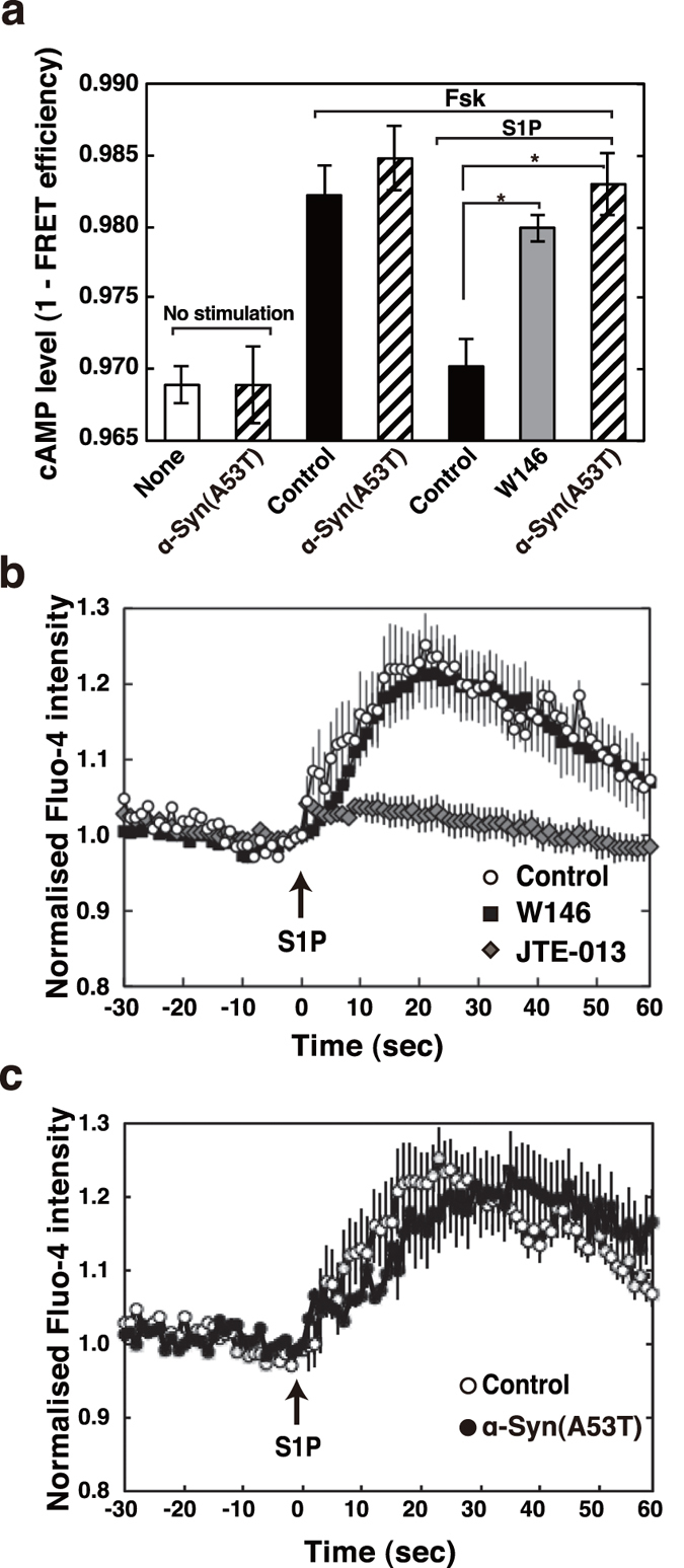
Figure 3. α-Syn-induced impairment of S1P1 receptor- but not S1P2 receptor-mediated signalling assessed in an endogenous protein system.

(a) SH-SY5Y cells transiently expressing the cAMP biosensor Epac1-camps were treated with 0.5 mM cAMP phosphodiesterase inhibitor, isobutylmethylxanthine, 20 μM forskolin with or without 100 nM S1P, or 10 μM W146 as indicated. Alternatively, cells transiently expressing the cAMP biosensor, which had been treated with 1 μM α-Syn(A53T) for 18 hr, were stimulated with each agonist as indicated (hatched bars). The FRET efficiency was estimated using acceptor photobleaching. Values represent means ± s.e.m. of 3 independent experiments carried out in triplicate. Statistical significance was analysed by Student’s t-test (*P < 0.05). (b) SH-SY5Y cells were serum starved for 18 hr and loaded with 2 μM Fluo-4 AM for 20 min. Cells were washed and pretreated with 10 μM W146 or 10 μM JTE-013 or without (control) for 10 min. The Fluo-4 emission signal for each cell was acquired at a frequency of 1 Hz by fluorescence microscope. After taking basal level signal 100 nM S1P was added (arrow) and the change in fluorescence was monitored. One of the representative quantification results of fluorescence changes in 40 control, 40 W146- and 40 JTE-013-treated cells from 3 independent experiments is shown. (c) SH-SY5Y cells were serum starved for 18 hr with or without 1 μM α-Syn(A53T). Cells were then loaded with Fluo-4 AM and stimulated with 100 nM S1P (arrow) in the absence or presence of 1 μM α-Syn(A53T). One of the representative quantification results of fluorescence changes in 40 control and 40 α-Syn(A53T)-treated cells from 3 independent experiments is shown.
