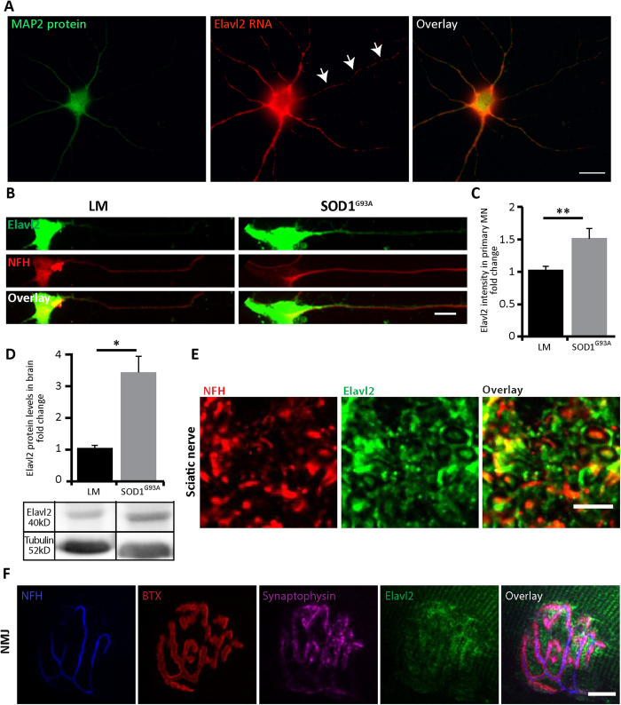
Figure 9. In-Vitro and In-Vivo Characterization of Elavl2 Expression in MN.
(A) In situ hybridization for Elavl2 mRNA in primary MN cultures from wild-type mice shows abundant mRNA in the soma and MAP2-positive dendrites. In addition, distinct puncta are present along the MAP2-negative axon shaft (arrows). Scale bar: 20 μm. (B) Immunofluorescence staining for Elavl2 in primary MN cultures from SOD1G93A and LM mouse embryos show increase in the axonal expression of Elavl2 in SOD1G93A MN. Green color indicates Elavl2, red color indicates NFH Scale bar: 10 μm. (C) Quantification of Elavl2 immunofluorescence intensity in axons only reveals a significant ~1.5 fold increase in the protein levels of Elavl2 in axons of SOD1G93A over LM controls. **p < 0.01 (student’s t-test; n = 61). (D) Western blot analysis of Elavl2 levels in brains of postnatal day 120 (P120) SOD1G93A and LM mice shows a significant increase in Elavl2 levels. Tubulin was used as loading control. *p < 0.05 (Student’s t-test, n = 3). The blot was cropped, and its full length is presented in Supplementary information. (E) Whole mount immunofluorescence staining of gastrocnemius muscle from P60 SOD1G93A for Elavl2 protein reveals enrichment its levels at the neuromuscular junction region, as shown by its co-localization with pre- and post-synaptic markers. Blue color indicates NFH in presynaptic MN. Red color indicates BTX staining for postsynaptic AchR aggregation. Green color indicates Elavl2. Magenta color indicates Synaptophysin in the presynaptic MN. Scale bar: 10 μm. (F) Immunofluorescence staining for Elavl2 in sciatic nerve cross-sections shows the presence of Elavl2 protein in axons. Red color indicates NFH, Green color indicates Elavl2. Scale 10 μm.

