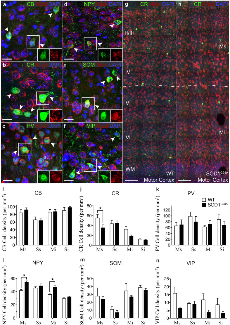
Figure 2. Calretinin and Neuropeptide Y interneuron subtypes are differentially altered in specific lamina of the SOD1G93A motor cortex.
(a–f) Calcium binding proteins and neuropeptides (green) were used to visualise specific interneuron populations in the cortex, showing labelling patterns of calbindin (CB; a) calretinin (CR; b) parvalbumin (PV; c) and neuropeptide Y (NPY; d) somatostatin (SOM; e) and vasoactive intestinal peptide (VIP; f) populations in 20 week WT cortex stained with DAPI (blue) and Nissl (red). The boxed areas (a–f) in the high magnification images show co-localisation of interneuron labels with Nissl stain. (g,h) At 20 weeks, analysis of motor cortex, reveals the normal distribution of CR-interneurons in WT motor cortex (g) but a striking reduction in particular in layers I-IV of SOD1G93A motor cortex. (h) Analysis of immunopositive neurons within the SOD1G93A motor (M) and somatosensory (S) cortex showed that the density of calretinin-expressing interneurons was significantly decreased specifically within the supragranular (Ms, layers I-IV) lamina of the motor cortex (j) and the density of Neuropeptide Y-expressing interneurons was significantly increased in both the supragranular (Ms, layers I-IV) and infragranular lamina (Mi, layers V-VI) of the motor cortex. (l) No other interneuron populations were significantly altered in either motor or somatosensory cortex. (i,k,m,n) Values in graphs represent means ± SEM. *P < 0.05, two-way ANOVA, Bonferonni’s multiple-comparison test with n = 6 mice per group. Scale bar in (a–f) 20 μm, (g–h) 100 μm.

