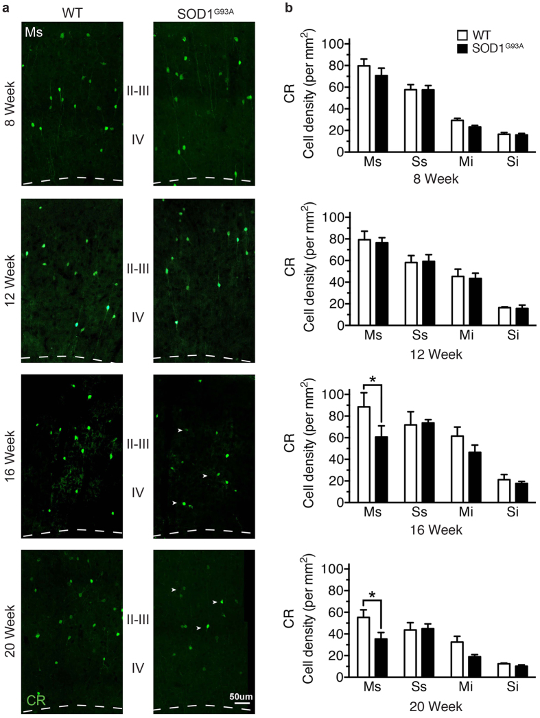
Figure 3. Calretinin-expressing interneurons are progressively lost during the symptomatic phase in the SOD1G93A motor cortex.
(a) CR-expressing neurons were labelled throughout the SOD1G93A disease course, showing neurons were present at comparable levels in SOD1G93A and WT mice at 8 weeks (early symptom onset) and 12 weeks in motor (M) and somatosensory cortex (S). (b) Analysis of 16 week symptomatic SOD1G93A mice showed that CR neurons were significantly decreased in the supragranular lamina of motor cortex (Ms, layers I-IV) compared to WT mice. CR-neurons were progressively reduced in the supragranular lamina of motor cortex (Ms, layers I-IV) in 20 week end-stage SOD1G93A mice (arrow heads in a). Values in graphs represent means ± SEM. *P < 0.05, two-way ANOVA, Bonferonni’s multiple-comparison test with n = 6 mice per group. Scale bar in (a) 50 μm.

