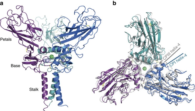
Figure 2. 3D structure of homo-CPI.
(a) View from the side showing the stalk, base and petal regions. Each chain is represented in a different colour, with bound Ca2+ ions as light blue spheres and the Cl− ion in green. Disulfide bonds are in yellow. (b) View from the top showing a structural alignment of the homo-CPI structure (in colour) on the 3.3 Å structure of CPIII (PDB code 4AK3; in grey). While overall the two structures are well aligned, there is a shift in orientation of helix-4 (highlighted for one chain from each structure).

