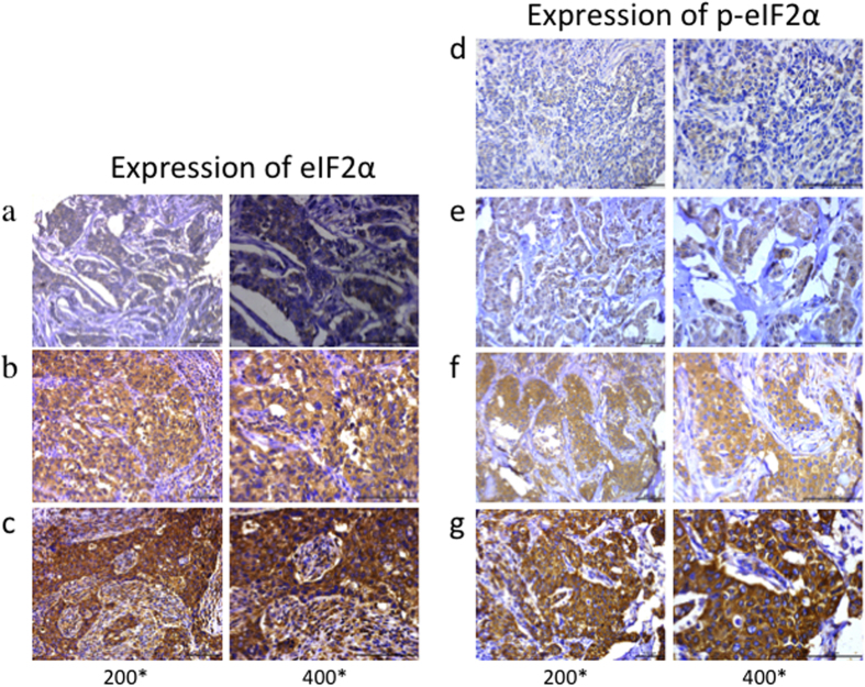
Figure 1. Total eIF2α and p-eIF2α expression profiles and scoring in breast cancer tissues.
Total eIF2α was evaluated in breast cancer and divided into three groups. Representative images of tissue scored as (a) 1, (b) 2, and (c) 3. p-eIF2α immunostaining was divided into four groups. Representative images of tissue scored as (d) 0, (e) 1, (f) 2 and (g) 3. Left photomicrographs, 200× magnification; right photomicrographs, 400× magnification.

