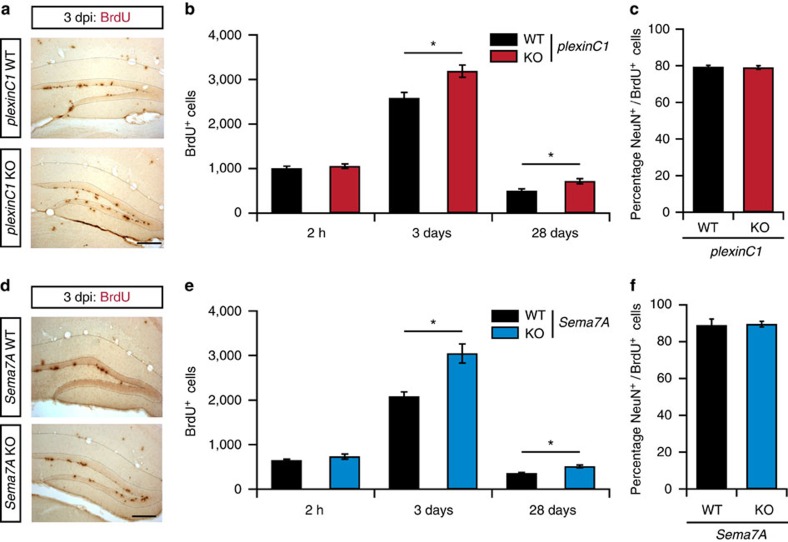
Figure 3. PlexinC1−/− and Sema7A−/− mice display increased cell proliferation in the adult DG.
(a,d) Representative examples of BrdU immunostaining in the dentate gyrus (DG) of adult wild-type littermates (WT), plexinC1−/− or Sema7A−/− mice at 3 days after BrdU injection (dpi). KO, knockout. (b,e) Adult mice were injected with BrdU and processed for immunohistochemistry for BrdU at 2 h, 3 days and 28 days post-injection. An increase in the number of BrdU-positive cells was detected at 3 and 28 dpi in plexinC1−/− and Sema7A−/− mice as compared to WT (two-way ANOVA, post-hoc t-test, 2 h: n(WT, plexinC1−/−)=4, n(Sema7A−/−)=3, 3 days: n(WT, Sema7A−/−)=4, n(plexinC1−/−)=7, 28 days: n(plexinC1−/−,-WT)=5, n(Sema7A−/−)=4, n(Sema7A-WT)=3). (c,f) Quantification of the number NeuN/BrdU-positive cells in the indicated mouse models at 28 dpi (Wilcoxon-Mann-Whitney test, Sema7A: n(Ki-67, DCX)=4, plexinC1: n(WT: Ki-67, DCX)=3, n(plexinC1−/−: Ki-67, DCX)=4, P(plexinC1)=0.8728, P(Sema7A)=0.7114). Data are presented as means±s.e.m., **P<0.01, *P<0.05. Scale bars: 250 μm.

