Fig. 4.
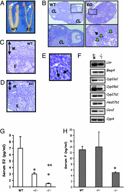
Female reproductive phenotypes. (A) Morphological analysis of the female reproductive tract indicates hypoplastic uteri and ovaries in the mutant (KO, Right) compared with those in a WT control (Left) at 9 weeks. White arrows denote ovaries. (B) Low-power histology of the ovary shows many corpora lutea (CL) in the WT (Left) but not in the mutant (KO, Right). Multiple follicles at different stages are present in the mutant section, but preovualtory follicles are not present (Right Upper). (Left Lower) A magnified region (rectangle) of the WT section that contains CL, an antral follicle with a healthy oocyte. (Right Lower) A magnified region (rectangle) of the KO ovary section with a follicle containing a collapsed oocyte (black arrow) and many degenerating follicles with remnants of zona pellucida (green arrows). (Magnification: ×5.) (C and D) Histology of a 9-week-old null (KO, D) mouse uterus shows a thin myometrium (M) and hypoplastic endometrium (E), compared with that from a control (WT) mouse (C). The vertical doublehead arrows indicate the relative thickness of the myometrium in both cases. (E) High-power image of a follicle from KO ovary shows theca (white arrow) is formed in the absence of LH. Black arrows indicate granulosa cells for reference to the interior of the follicle. (Magnification: ×20.) (F) RT-PCR assay indicates that thecal cell markers LH receptor and BMP4 are expressed in the null (–/–) ovary. Many steroidogenic pathway enzymes and cycloxygenase 2 are suppressed in the null ovary in the absence of LH. Lhr, LH receptor; Bmp4, bone morphogenetic protein 4; Cyp11a1, P450 side chain cleavage enzyme11a1; Cyp19a1, cytochrome P450 aromatase; Cyp17a1, cytochrome P450 17-α hydroxylase; Hsd17b1, 17-β dehydrogenase type I; Cox2, cycloxygenase 2. (G and H) Consistent with the ovarian and uterine phenotype, serum estradiol (E2; G) and progesterone (P; H) are suppressed in the null mutants (–/–), but only E2 levels are suppressed in the heterozygous mutants (+/–). *, P < 0.05 vs. WT; **, P < 0.05 vs. ±.

