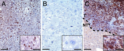
Fig. 5.
p53 immunostaining of Apc–/– HCC. (A) p53 accumulation in a hyperplastic liver of large T antigen-expressing ATIII-SV40 transgenic mice (37), used as a positive control for nuclear staining. (B) No p53 staining was seen in an Apc–/– MD HCC. (C) Intense p53 staining was seen in the nuclei of tumoral cells from an Apc–/– PD HCC. (Scale bars: 50 μm; 20 μmin Insets.)

