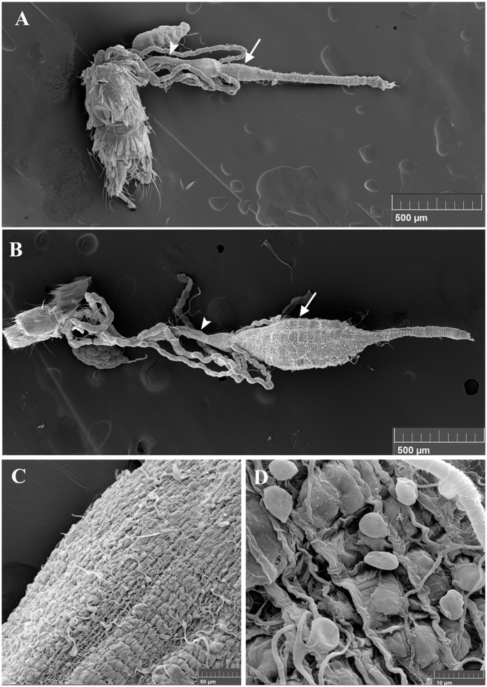
Figure 1. Scanning Electron Microscopy (SEM) images of A. albopictus mosquito guts.
The ultrastructure of the gut of a male (A) and a female (B) as revealed by SEM shows two of the three main compartments: the hindgut (arrowhead) with the associated malpighian tubules and the midgut (arrow). (C and D) are close-up photos of female guts where individual visceral muscle cells as well as tracheal cells and tubing surrounding the gut are visible.

