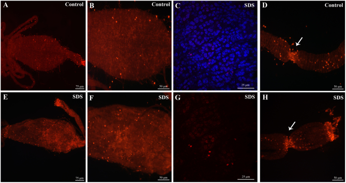
Figure 3. Regenerative cells are present in the midgut of adult A. albopictus.
Immunofluorescence staining using anti-PH3 antibodies reveals the presence of cells undergoing division in the gut of adult mosquitoes. An increase in the number of proliferating cells in the midgut is observed 24 hours after feeding the mosquitoes on SDS-sucrose (E and F) as compared to the midguts of control mosquitoes (A and B). A higher magnification picture focused on midgut cells labeled with anti-PH3 and DAPI staining is shown in (C and G). Two zones of active cell division (arrows) are observed in the most anterior part of the midgut independently of gut damage (D and H).

